The term ESD, which stands for Endoscopic Submucosal Dissection, refers to a technique for submucosal dissection via endoscopy. This is a new technique used for the treatment of early gastrointestinal cancer.
1. What is ESD?
Gastrointestinal cancer is one of the most common cancers, accounting for the largest proportion of various types of cancer. According to the latest research, the rate of gastric cancer in Vietnam ranks 18th among the 20 countries with the highest rates of gastric cancer worldwide. This alarming figure indicates that most patients are diagnosed at an advanced stage. However, if detected early, it is entirely possible to cure gastrointestinal cancer using the endoscopic mucosal resection method.
Currently, with the advancement of endoscopic submucosal dissection (ESD) for the treatment of early gastrointestinal cancer, early diagnosis of gastrointestinal cancer plays an extremely important role. In cases of severe dysplasia, precancerous conditions, or cancer confined to the mucosal layer of the gastrointestinal tract, if submucosal dissection is performed, the disease can be completely cured, and the patient’s lifespan can be extended to that of normal individuals.
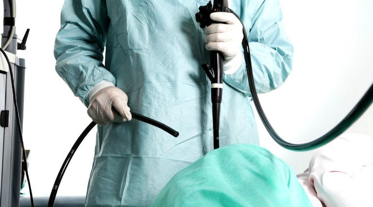
The endoscopic submucosal dissection (ESD) method is a minimally invasive procedure for treating early gastrointestinal cancer, helping patients preserve their entire stomach.
This method has been developed to address the limitations of traditional mucosal resection, with advantages such as minimal trauma, preservation of the digestive tract, improved quality of life for patients, and allowing patients to be discharged and continue regular medication just a few days later, with endoscopic lesions healing.
2. Some basic structural and functional characteristics of the digestive tract
The basic structure of the walls of the digestive tract consists of 4 layers, from inside to outside, including:
- Mucosa
- Submucosa
- Muscle layer (inner circular muscle and outer longitudinal muscle)
- Outer layer
- The digestive tract serves both the function of transporting and containing food, as well as the functions of digestion and absorption of nutrients found in food. Excess food waste is then concentrated and expelled through the anus.
- In the mucosa of the digestive tract, there are digestive glands. Depending on the specific structure and function of each segment of the digestive tract (esophagus, stomach, small intestine, colon, and rectum), the surface epithelial cells along with the digestive glands in a specific location have their own characteristics.
- The submucosa is connective tissue containing blood vessels, nerves, and the lymphatic system that support nourishment and the expulsion of toxins.
3. Indications and contraindications
3.1 Indications
- Lesions of differentiated adenocarcinoma, non-ulcerated type regardless of size, or ulcerated type adenocarcinoma with a diameter of less than 3cm.
- Undifferentiated cancer lesions, non-ulcerated, with a diameter of less than 2cm.
3.2 Contraindications
- Undifferentiated cancer lesions with a diameter >2cm, or differentiated cancer lesions with surface ulceration and a diameter >3cm.
- Suspicion of acute coronary syndrome.
- Uncontrolled hypertension.
- Suspicion of hollow organ perforation.
- Suspicion of aortic aneurysm or dissection.
- Patients in respiratory failure.
- Patients with severe heart failure.
- Patients with uncooperative psychiatric disorders.
- Relative contraindication: Systolic blood pressure < 90mmHg.
4. Advantages and disadvantages of the technique
- If resected below the mucosa, the disease can be completely cured, and the patient’s lifespan can be extended like that of normal individuals.
- Minimal trauma; preservation of the digestive tract.
- Improves the quality of life for patients.
- Can obtain lesions for pathological examination.
- The technique is difficult, requiring the endoscopist to have experience and precision in every step.
5. Procedure
- Step 1: Perform endoscopy according to standard procedures, carefully observing the lesion that needs to be resected, determining the boundaries of the lesion, and starting to mark the area to be dissected about 3-5mm from the boundary of the lesion.
- Step 2: Inject submucosally to elevate the mucosal layer containing the cancerous lesion.
- Step 3: Create a cutting margin around the lesion.
- Step 4: Dissect the submucosal layer, removing the lesion from the digestive tract.