Abnormalities in the breast that are too small to be detected by palpation can be identified through a breast biopsy. Ultrasound guidance plays a crucial role in early detection of breast cancer or other breast-related conditions.
1. Definition of Ultrasound-Guided Breast Biopsy
For abnormalities in the breast that are too small to be felt, an ultrasound-guided breast biopsy is performed. This procedure involves extracting a small sample of cells from the suspicious area for microscopic examination to determine whether they are benign or malignant.
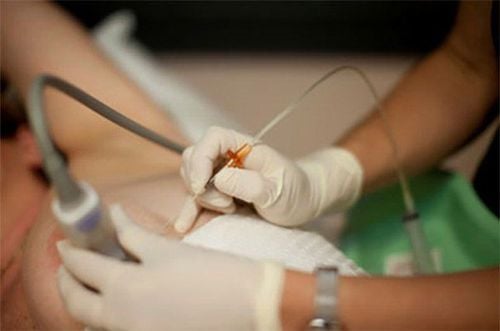
During the procedure, doctors use an ultrasound probe in one hand and a biopsy needle in the other. The ultrasound device provides real-time imaging to guide the needle precisely to the abnormal tissue. This ensures accurate sampling while minimizing scarring or cosmetic concerns.
2. Indications for Ultrasound-Guided Breast Biopsy
After undergoing clinical examinations and imaging tests such as breast ultrasound and mammography, a biopsy may be recommended if the following abnormalities are detected:
- A suspicious solid breast lump;
- Structural abnormalities in breast tissue;
- A region of abnormal tissue suspected of being breast cancer.
Additionally, an ultrasound-guided breast biopsy is indicated in cases such as:
- Lesions classified as BIRADS 4 or 5;
- Suspicious axillary lymph nodes;
- Recurring lesions or axillary lymph node abnormalities;
- Cystic lesions suspected to be malignant;
- Fluid-filled lesions requiring aspiration before biopsy.

3. Ultrasound-Guided Breast Biopsy Techniques
3.1 Core Needle Biopsy (CNB)
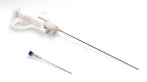
Core needle biopsy under ultrasound guidance is commonly performed for lesions categorized as BIRADS 4 or 5. This method collects tissue samples using a biopsy needle (typically 14G, about the size of a toothpick). Typically, five core samples are extracted. If the samples are insufficient, additional specimens may be taken.
With ultrasound guidance, the physician uses a specialized biopsy needle with an outer and inner core to cut and retrieve tissue samples efficiently while minimizing damage to surrounding tissue. The extracted tissue is preserved in formalin and subjected to histological staining for evaluation. This helps determine whether the lesion is benign or malignant, assess its severity, and classify cancerous conditions.

3.2 Large-Core Needle Biopsy with Ultrasound or X-ray Guidance
For smaller lesions, particularly microcalcifications, or cases where core biopsy samples are insufficient, a larger needle biopsy may be used. This method employs a vacuum-assisted large-core needle (up to two-thirds the size of a chopstick) to obtain substantial tissue samples for accurate diagnosis.
If the abnormality is visible only on mammography, X-ray guidance is used. However, if it is detectable on both mammography and ultrasound, ultrasound is preferred due to its flexibility and safety.
Because a larger needle is used, bleeding risk must be considered. Patients undergo blood count and coagulation tests before the procedure. A special anesthetic is also administered to minimize bleeding, and post-procedure compression is applied to the breast tissue.
This advanced method requires highly trained physicians and technicians. Patients receive thorough explanations regarding the procedure, its duration, and potential complications.
4. Advantages of Ultrasound-Guided Breast Biopsy
Breast ultrasound helps detect abnormalities and suspected tumors. If necessary, an ultrasound-guided breast biopsy is performed to differentiate between cystic and solid masses.
Additionally, ultrasound can identify small lesions that are undetectable by palpation or visual inspection. It is particularly useful for detecting abnormalities in pregnant women, as it is a safe diagnostic tool compared to other imaging techniques.
Ultrasound is especially effective for identifying solid breast masses in women over 35 years old, particularly when mammography results are inconclusive. By analyzing six morphological characteristics of a solid breast mass, doctors can determine whether the tumor is benign or malignant. These characteristics include:
- Shape;
- Orientation;
- Margins;
- Boundary definition;
- Internal structure;
- Acoustic shadowing.
Furthermore, ultrasound can detect early signs of cancerous changes, such as skin thickening over the breast. In such cases, an ultrasound-guided breast biopsy is typically recommended.
Ultrasound guidance ensures precise needle placement for biopsy, reducing patient discomfort and avoiding excessive pressure on the breast compared to mammography-guided procedures.

