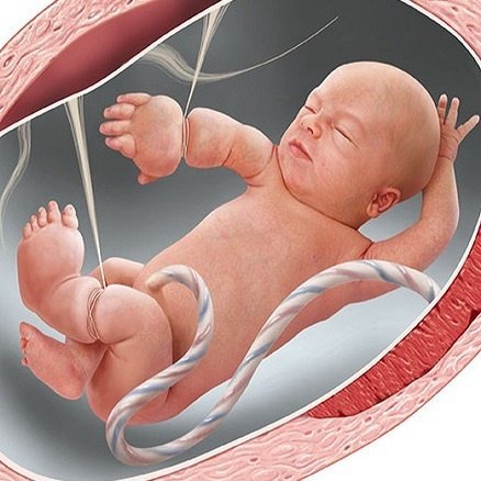
Amniotic bands are a congenital abnormality, occurring in a prevalence rate from 1:12,000 to 1:15,000 children, with boys and girls at equal risk; it manifests in the limbs (legs, arms, and torso).
1. Risk factors for amniotic band syndrome
Mother smoking; living in high-altitude areas…
The cause of the disease is unclear, and the pathogenesis is believed to be due to rupture of the amniotic membrane in the early stages of pregnancy, leading to the development of multiple amniotic bands from the chorionic membrane that entangle the embryo or germ disc.
This restricts the blood vessels that nourish and create constrictive bands, affecting the structure of organs. Other mechanisms such as vascular disorders, mechanical disorders, genetic disorders, etc.
2. Signs and symptoms for early detection
From the fetal stage, it can be screened through ultrasound based on:
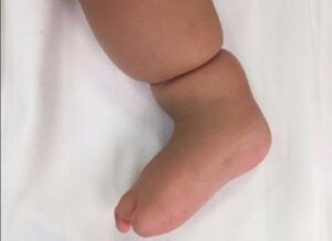
- Images of constrictive bands
- Deformities of the limbs: loss of distal parts of one or more fingers, syndactyly, limb absence, limb amputation, clubfoot…
- Abdominal wall deformities: chest abnormalities, abdominal hernia, scoliosis…
- Neurological defects: brain and spinal cord
- Craniofacial abnormalities: cleft face and skull.
3. Diagnosis of amniotic band syndrome
- Prenatal diagnosis: diagnosis in the first trimester, ultrasound can detect constricting rings, limb reductions, body wall defects, or craniofacial defects, with distal parts of the limbs showing swelling. An accurate prenatal diagnosis is often in severe cases: there are multiple major abnormalities as seen in limbs and the torso (short umbilical cord, amniotic bands). In the third trimester, 3D ultrasound or magnetic resonance imaging are supplementary techniques that can provide information to help determine the diagnosis. On T2 magnetic resonance images, the amniotic bands are observed as thin strips.
- Postnatal diagnosis: suspicion in children with limb reductions, trunk deformities, and cranial defects in unusual locations. Examination of the amniotic membranes and placenta is very important. Finding amniotic fibers will support the diagnosis.
- Differential diagnosis:
4. Prognosis of amniotic band syndrome
5. Management of amniotic band syndrome
- Prenatal assessment and counseling: Doctors should discuss with parents the potential risks that may occur during pregnancy and postpartum risks. Amniocentesis may be performed if it pertains to the decision to continue the pregnancy.
- Prognosis should be discussed with pediatricians, neurologists, and orthopedic surgeons. Some patients may have prenatal release of constriction bands.
- The release of constriction bands in the uterus must be carried out before severe and irrecoverable vascular damage occurs that affects the limbs, leading to amputation. The efficacy of this method has not been proven, nor is there consensus on the criteria for selecting patients for intervention. For amniotic bands related to a limb, a reasonable criterion is that the limb must have normal blood flow.
- Intervention in the fetus needs to weigh the benefits and risks. The frequency of prenatal monitoring or delivery methods does not have specific guidelines, depending on the injury. If blood vessels are constricted, fetal intervention should be considered.
- For newborns, some children with amniotic band syndrome may be mistaken for plump babies. Releasing the constriction bands reduces venous obstruction and lymphedema, which may save some limbs. Surgery may be delayed for weeks to months unless the case of vascular constriction requires emergency surgery. Additionally, children receive palliative care, including function-enhancing or cosmetic corrective surgeries.