
Accessory breasts (polymastia, supernumerary breasts, or mammae erraticae) is a condition where a person has at least one additional breast. Accessory breasts can appear with a nipple or areola. This is a relatively common congenital condition in which abnormal breast tissue appears alongside normal breast tissue.
1. Causes of accessory breasts
The condition of polymastia often occurs during the development in the fetus. Normally, breast tissue develops along the milk line, and the additional tissue dissolves and is absorbed by the body. Polymastia occurs when the additional tissues do not dissolve before birth. This condition can be hereditary.
2. Locations of accessory breasts
This normal variant can appear as a lump anywhere along the trajectory of embryonic breast tissue (from the armpit to the groin). Common locations include the armpit (60-70%), chest wall, vulva, knee, lateral thigh, buttocks, face, ear, and neck.
3. Manifestations of accessory breasts
This condition occurs in 2-6% of women and 1-3% of men. Although accessory breasts have existed since infancy, most people are unaware of them until puberty. Most women only discover they have accessory breasts when they are incidentally found on ultrasound or mammography.
Common manifestations of accessory breast tissue include discomfort, pain, milk secretion, axillary thickening, and localized skin irritation. Accessory breast tissue responds to hormonal stimulation and may become more prominent during menstruation, pregnancy, or breastfeeding. The size ranges from a small lump to a complete structure (which may include a nipple and areola).
4. Pathology of accessory breast tissue
Medical literature worldwide has described both benign and malignant pathologies of accessory breast tissue in adult women. Benign pathologies include fibroadenomas, mastitis, or fibrocystic changes. Accessory breast tissue tends to have a higher malignant transformation rate and occurs at an earlier age.
However, it is reassuring that breast cancer in accessory breast tissue has an incidence rate of only about 6%. The most common pathology is invasive ductal carcinoma, accounting for approximately 50% – 75%. Macromastia may occur during pregnancy as well as in adolescence.
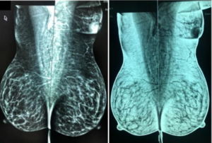
5. Imaging characteristics and examination methods
Most accessory breast tissue is found incidentally on routine mammography. Ultrasound imaging shows breast tissue that cannot be distinguished from normal breast tissue. Occasionally, magnetic resonance imaging of the breast is performed in atypical cases, exhibiting signal characteristics and enhancement similar to normal glandular tissue.
6. Treatment
Most cases of accessory breast tissue do not require treatment. However, when symptoms are present, the preferred treatment method is surgical excision, as it will reduce physical discomfort or mechanical discomfort in cases where the breast tissue is large.
REFERENCES
- Ersilia M. DeFilippis1 and Elizabeth Kagan Arleo, “The ABCs of Accessory Breast Tissue: Basic Information Every Radiologist Should Know”, American Journal of Roentgenology. 2014;202: 1157-1162. 10.2214/AJR.13.10930.
- Bickle I. and Radswiki, Assessory breast tissue, https://radiopaedia.org/articles/accessory-breast-tissue.
- Ruqayya Naheed Khan et al, “Invasive carcinoma in accessory axillary breast tissue: a case report”, Int J Surg Case Rep, 2019, 59: 152-155.